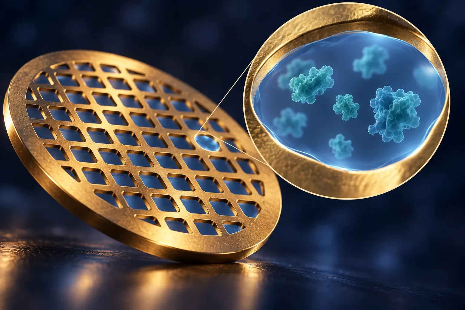
Cryo-electron microscopy can reveal extraordinary molecular detail. Its most consequential decisions often happen in a thin film, long before a polished structure appears.
The public image of cryo-electron microscopy is usually a finished molecular structure: a ribbon curling through a translucent envelope, a channel opening across a membrane, or a pocket that seems ready to receive a drug. The experiment begins with something less cinematic. A small amount of liquid must become a thin, frozen specimen that contains useful particles in useful arrangements. The quality of that transition can determine whether an expensive microscope records information or merely accumulates files.
On September 25, 2026, Zheng and colleagues reported a way to control specimen ice thickness using deliberately shaped graphene reservoirs. Their study connected liquid-film confinement with more uniform frozen layers and demonstrated reconstructions for several macromolecular specimens. The result is a preparation advance, not a declaration that every protein can now be imaged at the same resolution. Its importance is that a physical feature of the support helps determine what information reaches the reconstruction software. Control of ice thickness in cryo-EM via confinement
That is a useful place to begin a tour of the method. Instead of starting with the final structure and asking how impressive it looks, begin with the specimen and ask what survives each transition. Which molecules remain interpretable after preparation? Which directions are represented in the images? How much exposure can provide information before damage changes the object? Which details recur independently, and which are supplied by a model?
The focus here is single-particle cryo-EM of biological macromolecules. The examples are original, simplified reasoning exercises rather than laboratory protocols. They explain why a headline resolution number is only one part of the result, and why improvements in specimen preparation can matter as much as faster computation.
The film is an environment, not an empty window
A thin film is often drawn as a transparent holder for particles. That picture can encourage the assumption that the particles simply remain wherever they happened to be in the original liquid. The assumption deserves testing. A confined specimen has surfaces and a preparation history; neither disappears because the final material is frozen.
Noble and colleagues examined particle locations using tomography across more than fifty grid-and-sample preparations in a 2018 study. They found extensive association with air–water interfaces in that investigated set and showed how three-dimensional characterization could reveal features hidden in ordinary projections. The study supports examining the prepared specimen rather than presuming a uniform distribution. Its reported prevalence should not be treated as a universal percentage for every preparation method or protein. Routine single-particle sample and grid characterization by tomography
An original geometric thought experiment shows why surfaces become prominent. Consider a flat liquid slab with area A and thickness t. Ignoring its edges, its two broad surfaces have total area 2A, while its volume is At. The surface-area-to-volume ratio is therefore 2/t. Halving the thickness doubles that ratio. The arithmetic does not predict a particular protein's behavior, but it explains why a thin specimen cannot automatically be treated as a miniature version of a bulk solution.
A molecule near a surface may encounter conditions different from those in the liquid's interior. Whether that difference matters depends on the system. The important experimental question is not whether interfaces are intrinsically bad. It is whether the prepared population still represents the state relevant to the biological question.
This changes how a preparation improvement should be assessed. More particles per image might be useful, but it is not sufficient if the extra particles belong to a selected or altered population. A good comparison asks about particle condition, distribution, orientation and reproducibility, not only how densely a field of view is populated.
Thin enough for information, thick enough for the specimen
It is tempting to turn ice thickness into a competition for the smallest possible number. That framing misses the design problem. The specimen must accommodate the object and the measurement, and the useful thickness depends on what is being studied. An extreme that improves one aspect can worsen another.
Think of a hypothetical object whose largest dimension is comparable to the available film thickness. A preparation that looks favorable when judged only by the amount of surrounding material may constrain which orientations fit comfortably. A thicker region may accommodate more arrangements while introducing other imaging burdens. The optimum is therefore a relationship among object, support and acquisition, not a universal setting that can be copied between unrelated samples.
The 2026 confinement result makes the distinction between control and minimization especially helpful. Being able to choose and reproduce a thickness is more informative than occasionally finding a very thin patch. A reproducible variable can be compared systematically. A fortunate patch may deliver a structure without revealing why neighboring regions failed.
For a research program, that difference affects efficiency. If preparation conditions vary widely without being measured, each microscope session becomes a mixture of scientific investigation and troubleshooting. If the conditions can be characterized, the team can distinguish a specimen problem from an acquisition problem more quickly.
The reader should consequently ask two separate questions of a preparation claim. Did the method improve the best observed result? And did it make useful specimens more consistently? A single exceptional reconstruction can establish a capability. A distribution across independently prepared grids is needed to assess repeatability. The two achievements can arrive at different times, and neither should silently stand in for the other.
A projection does not reveal every arrangement
A two-dimensional image compresses information along the viewing direction. Two objects at different depths may overlap in projection; two different three-dimensional arrangements can produce similar views. Recovering a three-dimensional structure therefore requires more than enlarging a micrograph until its details become visible.
A simple paper exercise captures the issue. Draw two dots separated horizontally on a transparent sheet. Viewed straight on, the separation is obvious. Turn the sheet edge-on and the dots can overlap. Nothing about the objects changed, but the view lost the distinction. Repeatedly photographing the same edge-on arrangement does not restore the missing direction merely by increasing the number of photographs.
In single-particle analysis, many images contribute information about a structural population. Their usefulness depends partly on how their views complement one another. A collection with enormous redundancy in one view may constrain that view very well while leaving another aspect uncertain. The quantity of images and the diversity of informative images are related but different resources.
This is why a gallery of recognizable two-dimensional averages is encouraging but not conclusive. The gallery can reveal useful consistency, obvious contamination, or missing kinds of views. It cannot by itself guarantee a well-supported three-dimensional interpretation at every spatial frequency and direction.
For communication, an orientation distribution should be treated as part of the scientific result rather than as an obscure processing diagnostic. It helps explain where the reconstruction has strong support and where the data are comparatively thin. A reader does not need to understand every mathematical detail to appreciate the difference between many repeated views and a genuinely diverse set.
Tilting buys new views at a price

Original ideal-slab geometry: a tilted straight path has length t/cos(theta). Not an acquisition setting recommendation.
Tan and colleagues demonstrated a tilted-acquisition strategy for specimens with preferred orientations in a study published on July 3, 2017. Their work also used directional resolution analysis and examined practical consequences of tilt, including changes in effective thickness and imaging conditions. The demonstrations showed that new views could improve structural information, while the tradeoffs prevented a simplistic conclusion that a larger tilt is always better. Addressing preferred specimen orientation through tilting
The geometry can be understood with an idealized slab. If the slab's thickness perpendicular to its surface is t, a straight path through it at an angle θ from the surface normal has length t divided by cos θ. At zero degrees, the path is t. At sixty degrees, the cosine is one half and the path is 2t. This is a geometric illustration, not a recommendation for a microscope setting.
The example reveals the exchange. A changed view can supply information that the original orientation did not. It can also require the measurement to contend with a longer path through the specimen. The best choice depends on which limitation is currently most consequential.
An original decision comparison makes the point. Suppose a hypothetical dataset contains very clear views from one direction and almost none from another. Adding more equally clear views of the already dominant direction may improve precision without resolving the main ambiguity. Some less favorable but genuinely new views could contribute more to the structural question. That does not mean every low-quality tilted image should be kept. It means information value cannot be ranked by appearance alone.
A useful report separates these outcomes: changes in directional coverage, changes in overall resolution, changes in local interpretability and the costs of acquisition. Combining them into a single score can obscure why a method helped one specimen and disappointed on another.
The support can move even when the sample is cold
Freezing does not make the imaging problem mechanically motionless. Russo and Passmore's December 2014 study demonstrated gold specimen supports that substantially reduced support motion during irradiation relative to the compared carbon supports. Their work connected support stability with improved image quality and structural reconstruction, while also reporting residual motion. It is a historical experimental advance in the physical specimen platform, not a claim that modern images are free of movement. Ultrastable gold substrates for electron cryomicroscopy
The everyday analogy is taking a photograph through a moving window. A sharper lens cannot fully compensate for motion that smears the scene during the exposure. A later algorithm may estimate and correct some movement, but the ease of correction depends on what information remains in the recorded frames.
Now imagine two hypothetical acquisition strategies. One records a single integrated exposure. The other records a sequence of shorter frames whose total exposure is the same. If the object changes position during acquisition, the sequence may preserve clues about the trajectory that the integrated image conceals. The advantage is conditional: individual frames must still contain enough information for an appropriate correction, and the correction model must match the relevant movement.
This distinction prevents a common overstatement about computation. Correcting a known or estimable transformation differs from recovering information that was never recorded distinctly. An algorithm can use relationships across data, but it cannot make every loss reversible simply because it has many parameters.
The practical editorial question is therefore whether a reported improvement came from stabilizing the specimen, measuring its motion more effectively, correcting the recorded data more accurately, or selecting particles that happened to behave favorably. Each is a real route to progress. They imply different expectations for another sample or instrument.
Exposure is an information budget with a changing target
More illumination often improves an ordinary photograph. In biological electron microscopy, the object can change as it is measured, so extending exposure is not a cost-free route to detail. The useful question is how information and damage evolve together, not how bright an image can be made.
Nakane and colleagues' October 21, 2020, study demonstrated atomic-resolution single-particle reconstruction for apoferritin using advances in the electron source, energy filtering and detection. It also examined a membrane-protein receptor and discussed dose-related limits. The paper establishes striking performance for the investigated specimens and arrangements; it does not make its best resolution a guarantee for an arbitrary molecular complex. Single-particle cryo-EM at atomic resolution
An original statistical model illustrates one part of the tradeoff. Suppose independent observations of an unchanged signal contain zero-mean noise of the same variance. Averaging N such observations reduces the noise standard deviation in the mean in proportion to one divided by the square root of N. Four times as many observations gives half the standard deviation; sixteen times as many gives one quarter. These relationships require the stated assumptions.
If the underlying object changes during the measurements, averaging no longer estimates one unchanged object in the same simple way. Later observations can be more numerous and less representative of the state of interest. Likewise, if the noise is correlated, the improvement can be smaller than the independent-observation formula suggests.
This is why a scientific explanation should distinguish additional particles from additional exposure of the same particle. Both can increase the amount of recorded data, but they do not impose the same history on the specimen. Their usefulness also depends on whether the particles belong to the same structural population and whether their orientations can be estimated adequately.
The number of particles is not the entire sample size
A large particle count is easy to communicate. It is also easy to misinterpret. Some particles may contribute redundant views, some may represent different states, and some may have been recorded under conditions that provide less useful information for the question being asked.
Consider an explicitly fictional collection of one hundred thousand particle images. Suppose most belong to a common conformation, while a small fraction represent another state. An average of everything may emphasize the common structure and blur the differing region. The large total count does not mean that the rare state has been characterized with the same confidence as the common one.
Now suppose the team discards many images to obtain a sharper reconstruction of the dominant state. That can be entirely appropriate for a defined structural question. It also changes what the final map represents. The result is a structure of a selected population, not necessarily a description of every molecule in the original preparation.
The distinction becomes important when someone interprets the proportion of retained classes as a biological population measurement. Selection and classification can change those proportions. Without understanding those effects, a fraction in the processed dataset should not automatically become a fraction in the original solution or in a living cell.
A transparent account follows the population through the analysis. It distinguishes images acquired, particles proposed, particles retained, classes analyzed and final subsets used for particular maps. The purpose is not to celebrate a high retention rate. It is to make the relationship between the starting material and the reported structure intelligible.
Classification can clarify heterogeneity without proving a mechanism
The original cryoSPARC paper, published on February 6, 2017, introduced algorithms for efficient reconstruction and classification, including approaches that could generate initial structures without a supplied reference map. The accessible author abstract supports that historical algorithmic contribution; subscription methods were not inspected for this article. Its performance claims belong to the reported work, not to an assumed benchmark for current hardware or software versions. Punjani and colleagues, cryoSPARC
The underlying scientific issue is broader than a particular program. A mixed set of observations may contain several structures, continuous movement, measurement variation, or combinations of these. A classification algorithm imposes a way of representing that complexity. Its output is useful when the classes correspond to distinctions supported by the data, but the existence of several classes does not alone establish several biological mechanisms.
Imagine a hypothetical molecule with a flexible arm that moves continuously. A program asked to describe the data using three groups may return three representative arm positions. Those groups can help summarize the observations. They do not necessarily prove that the molecule occupies only three sharply separated states.
Conversely, two biologically meaningful states might be difficult to separate if the distinguishing feature is weak or poorly viewed. Failure to recover two classes would then be a statement about the analysis and data, not proof that the second state cannot exist.
A useful interpretation asks what alternative descriptions were tested and what features distinguish the selected groups. Repeated analyses with reasonable choices can reveal stability, but agreement among analyses sharing the same restrictive assumptions is not unlimited confirmation. The goal is to identify the structural variation the experiment genuinely resolves, then connect that variation to biology with additional evidence where needed.
Symmetry is powerful because it is a strong assumption
A symmetric object contains repeated relationships that can help an analysis. The same strength makes symmetry consequential. If an analysis imposes a symmetry that the relevant feature does not obey, the reconstruction can suppress or redistribute that feature.
An original toy example uses a ring with four nominally equivalent positions. Suppose a small attachment is present at only one position in each individual object. If the analysis averages all four positions as equivalent, the attachment's contribution can be spread across them. The resulting representation may show a weaker feature at each position rather than one fully occupied feature at a unique position.
This does not mean symmetric processing is improper. It means the question matters. If the goal is to characterize a symmetric core, the assumption may be useful and well supported. If the goal is to locate an asymmetric attachment or distinguish unequal subunits, the same assumption may hide the answer.
The interpretation also depends on whether the asymmetry is fixed relative to the object or varies across the population. Those situations can produce different biological implications even when a symmetric average looks similar. A final map should therefore be accompanied by enough information to understand which relationships were imposed and which emerged from observations.
For a reader evaluating a striking molecular image, a productive question is whether the feature carrying the main biological claim was examined under assumptions capable of preserving it. A high-resolution symmetric core does not automatically validate a low-occupancy asymmetric detail attached to it.
Independent halves test a particular kind of agreement
Scheres and Chen's 2012 study investigated overfitting in cryo-EM reconstruction and showed the value of independently refined halves of the data. Their tests contrasted apparently favorable resolution estimates with agreement to known structural references. The key contribution was a practical way to reduce misleading correlations caused by fitting noise. It should not be expanded into a claim that splitting data eliminates every preparation bias or modeling error. Prevention of overfitting in cryo-EM structure determination
The distinction can be explained with an original analogy. Two students independently measure the same object using the same misprinted ruler. Their answers may agree closely while sharing a scale error. Independence of their arithmetic does not remove the shared defect in the measurement system.
For a reconstruction, independent agreement is valuable evidence that particular details are reproducible within the data and processing arrangement. It is not evidence that every upstream assumption was correct. Both halves can contain the same selected population, the same missing orientations, or the same specimen changes.
The practical response is to ask what the validation test is designed to detect. A test for fitting independent noise should not be criticized because it does not answer a different question about biological representativeness. But it should not be advertised as answering that different question either.
This is a general feature of good validation: several tests can be complementary without being interchangeable. Internal consistency, agreement with independent measurements, chemical plausibility and repeatability across preparations each constrain different possible mistakes. Their combined value comes from preserving those distinctions rather than collapsing them into a single reassuring adjective.
Resolution varies across a structure
Kucukelbir, Sigworth and Tagare introduced a method for estimating local resolution in a paper first published online on November 10, 2013, and appearing in a 2014 issue. The accessible primary abstract reports analysis of spatially varying resolution across example datasets. Only the abstract and public metadata were reviewed here, so this article does not claim an independent assessment of its full algorithm. The work supplies a useful reminder that a whole-map resolution figure does not describe every region equally. Quantifying local resolution of cryo-EM density maps
Imagine a hypothetical complex with a rigid central body and a flexible extension. The central body may align consistently across particles, while the extension occupies several positions. An average can preserve detailed central features and leave a diffuse outer region. Reporting only the best-resolved part would hide the limitation precisely where a biological hypothesis might depend on the extension.
This is different from directional resolution. Local variation asks where information is stronger or weaker within the object. Directional variation asks whether different spatial directions are supported differently. A map can have both problems at once, and improving one does not necessarily improve the other.
For model interpretation, the relevant question is local and specific. Does the region support the proposed side-chain arrangement, small-molecule position or interaction? A detailed neighboring region does not lend its resolution to the uncertain feature. The evidence must be inspected where the claim is made.
A useful public illustration would show variation honestly instead of giving every part of the structure the same crisp rendering. A polished molecular model can otherwise make uncertain coordinates appear as definite as strongly supported ones. The style of the illustration should not erase the unevenness of the evidence.
The atomic model is another layer of interpretation
A reconstructed map and an atomic model are related objects, not synonyms. The map represents information inferred from images. The model assigns atoms and molecular geometry in a way intended to explain that information while respecting other constraints. A model can be chemically reasonable in a region where the map supplies only limited detail.
An original analogy is assembling a familiar machine from a partially obscured photograph. Prior knowledge helps identify plausible parts and arrangements. It can also encourage a person to complete an expected feature that the photograph does not distinguish. The finished drawing may be useful, but its most confident-looking lines need not all come from the image.
This distinction becomes especially important when interpreting a small feature. Suppose a hypothetical map supports an elongated region compatible with several chemical arrangements. A model-building procedure selects one because it fits prior expectations. The correct statement may be that the selected arrangement is consistent with the evidence, not that every alternative has been experimentally excluded.
Testing alternatives can strengthen the conclusion. If competing plausible models produce distinguishable predictions that the data favor differently, the ambiguity narrows. If several remain comparably compatible, reporting that range may be more informative than presenting one precise coordinate set without qualification.
There is no virtue in ignoring chemical knowledge. The point is to identify where that knowledge enters. An excellent model combines experimental evidence and well-founded constraints transparently. It becomes misleading only when the combined result is described as though every detail had been independently observed.
A pocket does not establish a drug's entire story
Molecular structures are often discussed through their potential relevance to medicines. A resolved pocket or a bound molecule can be important evidence about a physical interaction. It does not, by itself, establish therapeutic benefit, selectivity in a complex biological setting, or safety.
Consider a fictional binding study in which a molecule is present in a prepared protein sample. A structure may help explain how the molecule fits under those conditions. To connect that observation to a broader mechanism, investigators may need evidence that the interaction changes the relevant function. To connect it to treatment, further questions arise about exposure, competing interactions and biological outcomes. Those are separate experiments, not details supplied by a sharper picture.
Even the structural comparison requires care. If a ligand-bound and an unbound preparation differ in several conditions, an observed conformational difference cannot automatically be assigned solely to the ligand. The study design must make the attribution credible. A comparison between two beautifully resolved structures can still contain a confounding variable.
This article does not assess any medicine or propose clinical action. The point is about scientific communication: structural evidence should be described at the level it supports. Saying that a map suggests a binding interaction can be both exciting and accurate. Calling the same map proof of a treatment mechanism across an organism skips necessary steps.
The best use of a structure is often to generate a more discriminating next experiment. A predicted contact can be perturbed, a proposed functional change can be measured, or a competing interpretation can be tested. The structure gains explanatory power when those independent tests agree, not merely when its rendering becomes more elaborate.
A frozen population is not a movie of one molecule
Multiple structural states can suggest movement, but arranging them into a sequence does not automatically measure a trajectory. A collection of frozen configurations supplies information about the observed population under preparation conditions. Inferring how one molecule moves between states requires additional assumptions or time-resolved evidence.
An original thought experiment helps. Imagine photographs of different doors, some open and some closed. The collection demonstrates the existence of both configurations. It does not reveal how quickly a particular door opens, whether it pauses halfway, or which direction a photographed door was moving. A plausible animation can illustrate a hypothesis without becoming a measurement of motion.
The same distinction applies when an analysis produces a continuous-looking series of conformations. The series may summarize a structural landscape or a computational interpolation. Its ordering and spacing should not silently become time. Equal steps in an animation are not evidence of equal durations in a physical process.
This does not reduce the value of conformational analysis. Identifying distinct arrangements can constrain mechanisms and guide kinetic experiments. The communication challenge is to label what the visualization represents: observed classes, inferred variation, interpolated transitions or measured time points.
For a general reader, the most useful question is simple: did the experiment observe the same process at defined times, or did it arrange different structures into a possible story? Both can advance understanding. Confusing them makes the story seem more directly observed than it was.
Preparation repeatability can be tested as its own result
A method that produces one excellent grid has established an opportunity. A method that repeatedly produces useful grids under documented conditions has established something more operational. These are related achievements, but they require different evidence.
Consider a hypothetical comparison between preparation routes A and B. Route A yields one outstanding grid and nine poor grids. Route B yields eight useful grids with somewhat less spectacular best-case detail. Which route is preferable depends on the scientific goal, available sample and microscope time. The best resolution alone cannot answer the question.
An informative comparison would define useful before inspecting the results. The criterion might concern particle integrity, usable area, orientation diversity or suitability for a particular structural task. Defining the criterion afterward risks selecting whichever measure makes the preferred route look strongest.
The comparison should also distinguish independent preparation from repeated images of one successful grid. Many fields of view can characterize that grid thoroughly without demonstrating that the preparation procedure is repeatable. Different days, sample batches or operators can reveal variability that a single concentrated session does not.
Such tests need not be framed as bureaucratic obstacles. They identify where a method saves resources and where it remains sensitive. If a preparation route works well only within a narrow sample range, that knowledge helps laboratories choose it appropriately. A bounded, reproducible capability is more useful than an unrestricted promise supported by a few exceptional examples.
The bottleneck can move without disappearing
Progress in one stage changes which problem dominates next. Better preparation may expose a limitation in acquisition. Faster acquisition may shift effort toward computation or interpretation. Faster reconstruction may make validation and biological follow-up the most time-consuming tasks.
A simple original throughput example illustrates this. Suppose a fictional workflow requires ten hours for preparation, ten for acquisition and eighty for analysis, all performed sequentially. Halving the acquisition time reduces the total from one hundred hours to ninety-five, a five-percent reduction. Halving analysis reduces the total to sixty, a forty-percent reduction. The arithmetic says nothing about actual laboratory timings; it shows why a large improvement in one component may produce a modest end-to-end change.
Now suppose the stages overlap or share constrained equipment. The same arithmetic no longer captures throughput correctly. A queue for microscope access, limited sample availability or repeated failed preparations can dominate the schedule. A credible efficiency claim therefore identifies the workflow boundary and the resource being improved.
The useful endpoint is not necessarily structures per week. A program might care about independently validated answers to a biological question, successful characterization of difficult samples, or reliable comparison of multiple conditions. A pipeline optimized for the easiest benchmark can perform poorly against those objectives.
This suggests a practical way to read technology announcements. Ask which bottleneck was reduced, under what conditions, and what now limits the complete task. That question acknowledges genuine progress while keeping a component demonstration from becoming an unsupported claim of universal automation.
The final figure should retain the experiment's distinctions
A good final figure can carry much of this reasoning without becoming cluttered. It can distinguish a measured-image example, a reconstructed map, an atomic interpretation and an illustrative animation. It can identify where resolution varies and avoid assigning equal visual certainty to every modeled detail.
The caption is especially important when several layers appear together. A translucent envelope surrounding an atomic model can look like two independent confirmations even when the model was built directly against that envelope. Stating their relationship prevents a persuasive visual arrangement from exaggerating the amount of independent evidence.
Original conceptual art has a different role. It can make a thin specimen or a large instrument understandable at a glance, but it should not imitate an experimental micrograph without disclosure. Symbolic particles are useful for explanation precisely when nobody is asked to treat their shapes or positions as measured results.
For the technical record, retaining intermediate information matters more than making the public figure exhaustive. Other researchers may need the underlying images, processing choices, independent reconstructions and relevant preparation details to examine a disputed feature. A beautiful final model is not a substitute for the observations that constrain it.
The most persuasive story therefore allows the reader to move in both directions: from specimen to interpretation, and from a proposed biological claim back to the particular evidence supporting it. If that return journey stops at a polished picture, the explanation is incomplete.
Choosing the next comparison
Suppose a hypothetical team has enough material for only two further preparations. Its current reconstruction has a stable core and an ambiguous peripheral feature. One proposal is to repeat the same preparation and collect more images. Another is to change a preparation condition suspected of selecting against the peripheral arrangement. The right choice depends on the competing explanations, not on which proposal promises the larger file count.
If the ambiguity mainly reflects random measurement noise, more comparable observations may help. If the relevant configuration rarely survives the existing preparation, repeating it may reinforce the same selection. If the feature is genuinely flexible, neither strategy alone may produce one uniquely sharp arrangement. Writing these alternatives before collecting more data makes the experiment more informative.
The team can then specify what each outcome would mean. A feature that changes with preparation but leaves the core stable raises a different question from a feature that remains variable across all conditions. A result that appears only after a particular processing choice deserves a different investigation from one visible under several reasonable analyses. This is original experimental reasoning, not a reported outcome from the cited studies.
The value of the comparison is that it can change the next decision. It turns an ambiguous picture into a set of testable possibilities rather than treating ambiguity as a demand for unlimited additional acquisition.
What the ice makes possible
Cryo-EM's achievements are real. The method can produce extraordinary structural detail, and improvements in physical preparation, instrumentation and algorithms have expanded what can be investigated. The lesson of the thin film is not that those achievements should be distrusted. It is that their success depends on a chain of specific, testable conditions.
The next useful advance may look less dramatic than a new molecular rendering. It may be a support that behaves more consistently, a preparation that preserves a difficult population, or an acquisition strategy that supplies previously missing views. Those changes can alter the reliability of everything downstream.
For a reader, the strongest questions remain concrete. What population entered the analysis? Which views support the relevant feature? What does the validation test exclude? Is the biological claim about the whole structure, a well-resolved region, or a more uncertain interpretation? Where did prior knowledge help complete the model?
A finished structure is most valuable when those questions have clear answers. The frozen film then becomes more than a technical prelude. It is the first part of the evidence that allows the molecular picture to mean something.